digital media
Lecture on the fallopian tube
digital media
Virtual Hysteroscopy 1
digital media
Virtual Hysteroscopy 2
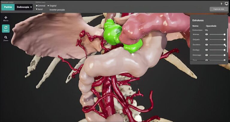
Medical imaging techniques, such as ultrasound (US) and magnetic resonance imaging (MRI), have been widely used to evaluate the uterine cavity and fallopian tubes (FT).

Uterine cavity malformations are congenital anomalies in the structure of the uterus that can impact fertility, increase the risk of miscarriage, and lead to pregnancy complications. These malformations result from defects in the development, fusion, or resorption of the Müllerian ducts during embryogenesis (Table 1). Diagnosis is performed through imaging techniques such as 3D ultrasound (US), hysterosalpingography, and MRI.

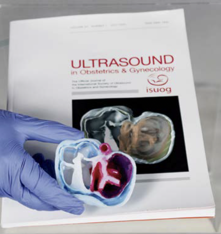
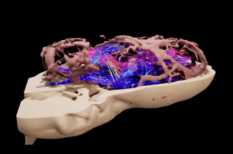
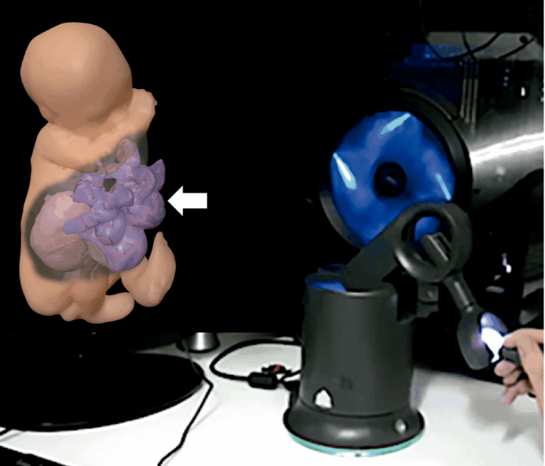
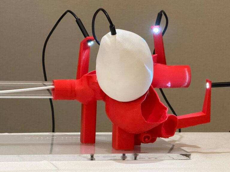
Hysteroscopy is considered the gold standard for assessing the endometrial cavity. However, it is an invasive and often uncomfortable procedure. Alternatively, contrast-enhanced US can also be used for this assessment. Three-dimensional hysterosalpingo-contrast sonography (HyCoSy), which incorporates second-generation contrast agents like SonoVue and advanced software in US machines, allows for a detailed evaluation of the uterine cavity and FT patency. This technique creates a Virtual Hysterosalpingography (VH), providing a non-invasive visualization of the uterine cavity and fallopian tubes.

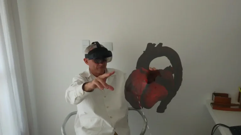
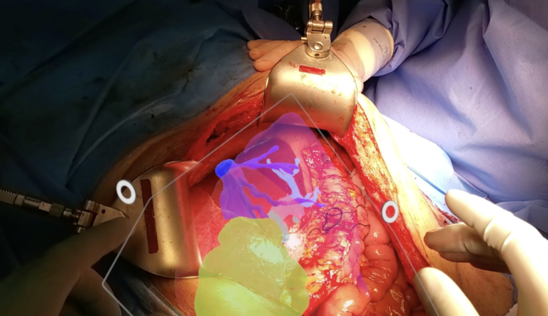
We believe that combining VH and Expanded Reality can offer valuable insights into the morphology of the uterine and fallopian tube cavities in addition to facilitating medical education and multidisciplinary discussions. However, further prospective studies are needed to confirm our findings.

Table 1: Types of Uterine Malformations
| Type of Uterine Malformation | Description |
|---|---|
| Septate Uterus | – A fibrous septum partially or completely dividing the uterine cavity. – The most common anomaly, frequently linked to recurrent miscarriages. – Can be corrected via hysteroscopic surgery. |
| Bicornuate Uterus | – Results from incomplete fusion of the Müllerian ducts, leading to two partially separated cavities. – Associated with an increased risk of miscarriage and preterm birth. |
| Unicornuate Uterus | – Occurs when only one Müllerian duct fully develops. – Leads to a smaller, asymmetrical uterus. – Can cause difficulties in conceiving and a higher risk of ectopic pregnancy. |
| Uterus Didelphys | – Develops when the Müllerian ducts fail to fuse, resulting in two separate uteri. – May be asymptomatic but increases the risk of pregnancy complications. |
| T-Shaped Uterus | – Associated with fetal exposure to diethylstilbestrol (DES) during pregnancy. – Characterized by a narrow uterine cavity, which can hinder embryo implantation and elevate the risk of miscarriage. |